

Poster no: 147
Poster no: 147
147
General anesthetics suppress behavior. While slow-wave power marks anesthesia, whether drug-specific mechanisms remain unclear. Traveling-wave analysis reveals cortical dynamics.
Methods
Multichannel electroencephalography (EEG) was recorded from two macaques during anesthesia sessions with propofol (n = 6), sevoflurane (n = 6), and esketamine administered on a background of sevoflurane anesthesia (n = 8). Behavioral arousal was scored from 0 to 4 using sequential responses to spontaneous movement, shaking/prodding, toe pinch, and corneal reflex. Spectral and traveling-wave analyses were performed. Traveling-wave dynamics were characterized along three dimensions: propagation extent, propagation order, and propagation pattern. Score- and condition-related effects were tested using linear mixed-effects models.
Results
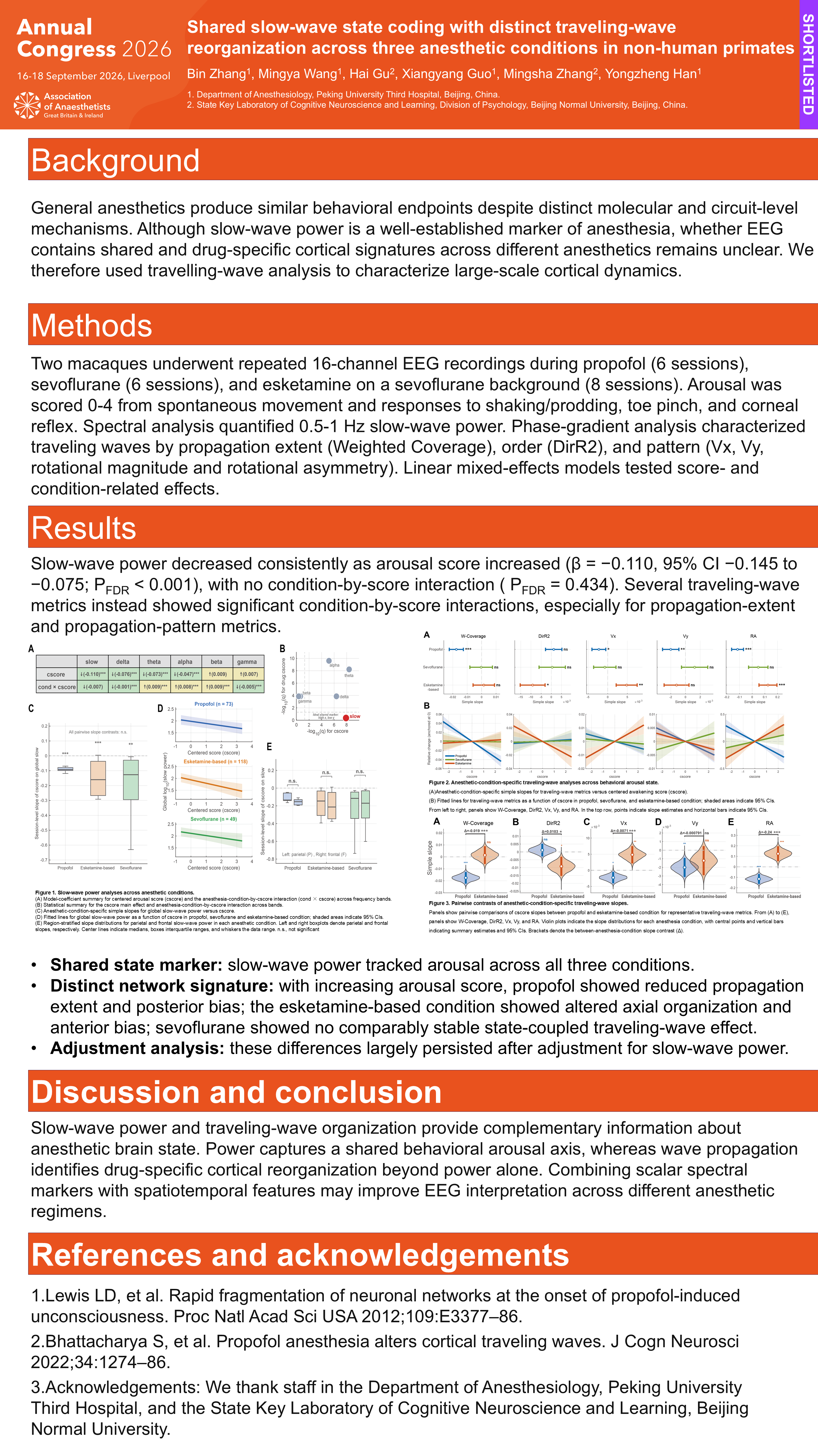
Across anesthetic conditions, slow-wave power showed the most consistent association with behavioral arousal score, decreasing as arousal score increased (β = -0.110, 95% CI [-0.145 to -0.075], P < 0.001). Slow-wave power also increased with within-drug anesthetic concentration (β = 0.133, 95% CI [0.068 to 0.199], P < 0.001), and this concentration dependence was stronger in segments adjacent to arousal score transitions than in behaviorally stable segments (Δβ = 0.158, 95% CI [0.056 to 0.259], P = 0.003). In contrast, slow-wave traveling-wave dynamics showed drug-by-score interactions, most prominently for propagation extent and topology metrics. Propofol was associated mainly with reduced propagation extent and posteriorly biased organization, whereas esketamine-based condition was associated with changes in axial organization and anteriorly biased organization; no comparably stable state-coupled traveling-wave effects were identified under sevoflurane. These traveling-wave

Figure 1. Drug-specific traveling-wave patterns across behavioral arousal state.
Fitted lines for traveling-wave metrics as a function of cscore in propofol, sevoflurane, and esketamine.
Discussion
Slow-wave power indexes a shared arousal axis[1] across propofol, sevoflurane, esketamine; travelling-wave patterns—independent of power—reveal drug-specific cortical reconfiguration[2] for EEG monitoring.
Acknowledgements
We thank staff in the Department of Anesthesiology, Peking University Third Hospital, and the State Key Laboratory of Cognitive Neuroscience and Learning, Beijing Normal University.