
Poster no: 141
Poster no: 141
141
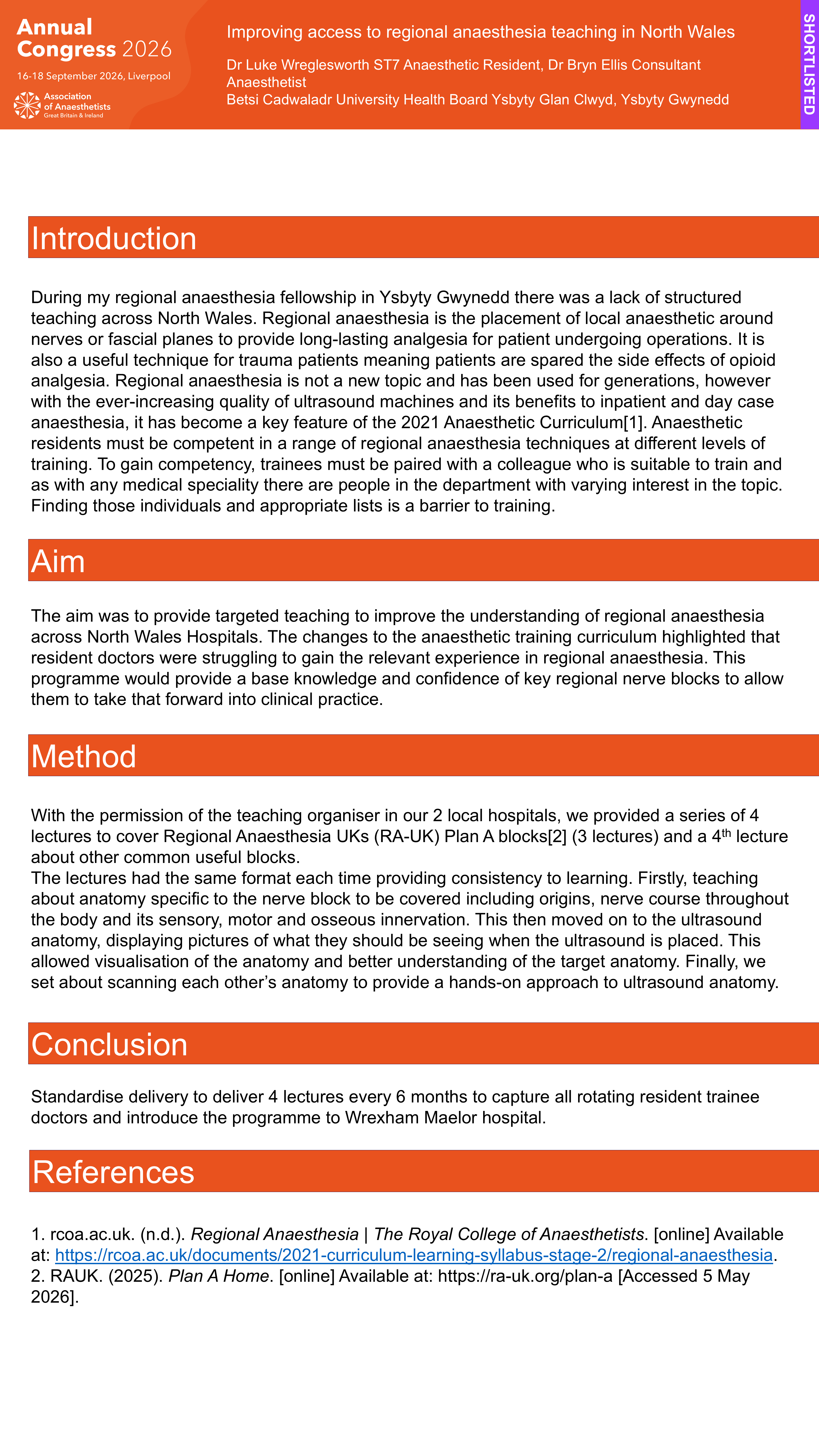
During my regional anaesthesia fellowship in Ysbyty Gwynedd there was a lack of structured teaching across North Wales. Regional anaesthesia is the placement of local anaesthetic around nerves or fascial planes to provide long-lasting analgesia for patient undergoing operations. It is also a useful technique for trauma patients meaning patients are spared the side effects of opioid analgesia. Regional anaesthesia is not a new topic and has been used for generations, however with the ever-increasing quality of ultrasound machines and its benefits to inpatient and day case anaesthesia, it has become a key feature of the 2021 Anaesthetic Curriculum [1]. Anaesthetic residents must be competent in a range of regional anaesthesia techniques at different levels of training. To gain competency, trainees must be paired with a colleague who is suitable to train and as with any medical speciality there are people in the department with varying interest in the topic. Finding those individuals and appropriate lists is a barrier to training.
The aim was to provide targeted teaching to improve the understanding of regional anaesthesia across North Wales Hospitals. The changes to the anaesthetic training curriculum highlighted that resident doctors were struggling to gain the relevant experience in regional anaesthesia. This programme would provide a base knowledge and confidence of key regional nerve blocks to allow them to take that forward into clinical practice.
Methods
With the permission of the teaching organiser in our 2 local hospitals, we provided a series of 4 lectures to cover Regional Anaesthesia UKs (RA-UK) Plan A blocks[2] (3 lectures) and a 4th lecture about other common useful blocks. The lectures had the same format each time providing consistency to learning. Firstly, teaching about anatomy specific to the nerve block to be covered including origins, nerve course throughout the body and its sensory, motor and osseous innervation. This then moved on to the ultrasound anatomy, displaying pictures of what they should be seeing when the ultrasound is placed. This allowed visualisation of the anatomy and better understanding of the target anatomy. Finally, we set about scanning each other’s anatomy to provide a hands-on approach to ultrasound anatomy.
Discussion
Standardise delivery to deliver 4 lectures every 6 months to capture all rotating resident trainee doctors and introduce the programme to Wrexham Maelor Hospital.